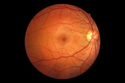
This technology uses high-resolution, digital photography to capture an image of the back of the eye. Capturing the image is as simple as looking into a camera an optician pushing a button, the image will be ready for immediate review by your doctor. The doctor will be able to see the retina, macula, optic nerve and blood vessels. The photo can show signs of macular degeneration, glaucoma, retinal holes, retinal detachments and high-blood pressure related eye conditions. The Optomap Retinal Image becomes a part of your records allowing the doctor to refer back to them each year.
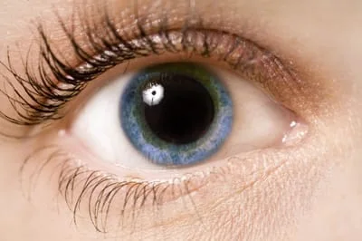
Dilation
This procedure uses drops to open the pupil so your doctor may see all around the inside of your eyes for detection of retinal disease, including retinal detachment. Drops may cause light sensitivity or blurred near vision. Talk to your doctor if you are concerned about the use of drops.